Digital dermatitis (DD), often referred to as hairy heel warts, is an infectious foot disease in dairy and beef cattle herds. It has been reported on 70 percent of all U.S. dairies and 95 percent of large operations comprising 500 or more cows. The size and scope of digital dermatitis within the beef industry is undetermined but is a growing concern. Once introduced to the herd, the disease can spread rapidly. It can often lead to lameness in cows, which decreases milk production and reproductive health (fertility) in dairy animals. In beef, lameness can account for 70 percent of all sales of non-performing cattle.
Management, Prevention and Trace Mineral Nutrition
From calves to heifers and lactating cows to dry cows, there is a need for an integrated prevention strategy that manages risk factors to help control digital dermatitis. This strategy should include the following:
Management
- Follow biosecurity measures to prevent infected animals from being introduced into the herd.
- Regularly inspect hind feet through digital dermatitis pen walks.
- Keep records of lesions on animals for informed decision-making based on their history.
- Identify M2 lesions early and treat promptly.
Hygiene
- Promote a clean, dry environment, and use footbaths as needed.
Nutrition
- Increase awareness of nutrition’s role in helping to prevent digital dermatitis during all cattle phases, including heifers and young stock.
- Formulate diets for adequate nutrient and micronutrient fortification.
- Availa® Plus, when fed as part of a specific DD formula and a well-balanced diet, has been shown to significantly decrease the prevalence and severity of digital dermatitis in non-lactating cattle.
Digital Dermatitis Identification and Disease Progression
An understanding of how to identify the presence and severity of digital dermatitis is key to keeping the disease under control. Once the disease is introduced into a herd, it spreads rapidly, and prevalence often exceeds 70 percent. There are five stages of digital dermatitis originally identified (Döpfer et al., 1997). The following guide will provide you with a visual of the disease (lesion) stage and its progression.
M0 — Healthy Claw
Disease Stage: Normal digital skin, no signs of dermatitis.
Progression: Common factors of an M0 turning into an M1 lesion are poor hygiene, sub-standard biosecurity measures and muddy pens.
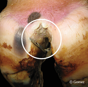
M1 — Early/Subclinical
Disease Stage: Small (less than 0.75 inch or 2 cm in diameter) circumscribed red to gray epithelial defects. Lesions can appear in the interdigital space. They can also occur between acute episodes of DD lesions or within the margins of a chronic M4 lesion as an intermediate stage.
Progression: An M1 lesion may develop into an M2 lesion or return to M0. Some animals with M1 lesions never develop M2 lesions.
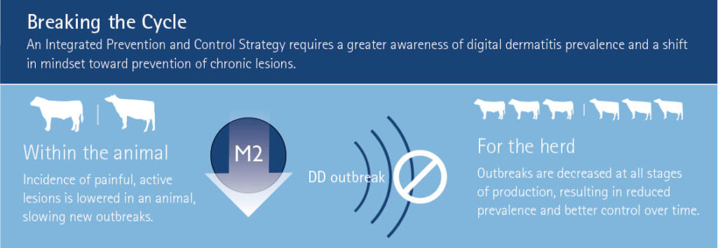
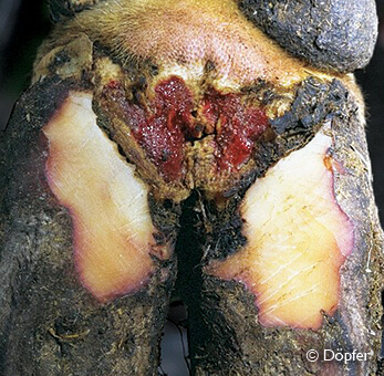
M2 — Painful/Acute Ulcer
Disease Stage: Bright-red active ulcer or red to gray granulomatous digital skin alteration greater than 0.75 inch (2 cm) in diameter. Commonly found along the coronary band at the skin/horn border, in addition to around the dew claws, in wall cracks and occasionally as a sole defect.
Progression: When treated effectively, healing (M3) begins. The lesion may cycle between M2 and M4.1 lesions. Accumulation of high numbers of M2 lesions starts an outbreak.
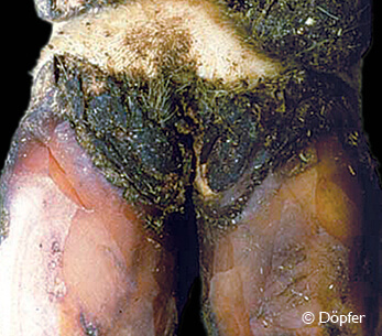
M3 — Healing
Disease Stage: Lesion surface becomes firm and scab-like within one to two days following treatment of an M2 lesion with topical antibiotics. Best-case scenario after topical therapy, the lesion appears to be no longer painful.
Progression: Healing can continue with lesion becoming M0. May regress to M2 or develop into a chronic M4 lesion.
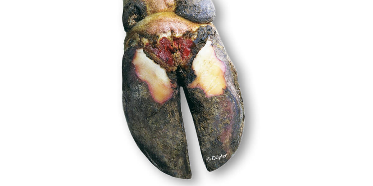
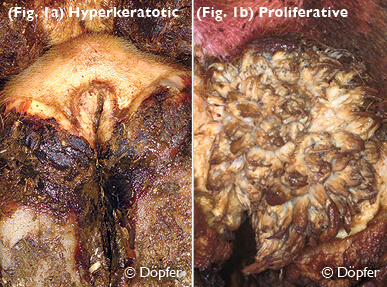
M4 — Chronic/Hairy Warts
Disease Stage: Rapidly growing hyperkeratosis (thickened epithelium; Fig. 1a) or filamentous, scaly or mass proliferations, commonly called hairy warts (Fig. 1b).
Progression: There is a reservoir of disease from encysted bacteria deep in the skin. Therefore, the lesion may return to M0 or progress to an M4.1 lesion.
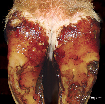
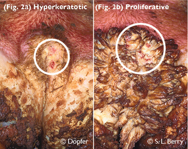
M4.1 — Chronic Recurring
Disease Stage: Hyperkeratosis and lesion within chronic M4 lesion (Fig. 2a) or chronic M4 lesion with an early or intermediate M1 lesion within its perimeter (Fig. 2b).
Progression: There is a reservoir of disease from encysted bacteria deep in the skin. Therefore, the lesion may return to M4 lesion or develop into M2 lesion.
For more information about how to assess, diagnose and prevent digital dermatitis in dairy cattle, download the Digital Dermatitis Field Guide or the Digital Dermatitis: Management and Prevention in the Feedyard guide for beef cattle.
Note: this article was developed in conjunction with Dr. Dörte Döpfer, University of Wisconsin School of Veterinary Medicine.